Scanning Electron Microscopy Processing unit
Transmission Electron Microscopy Processing unit
Histopathology Processing unit
Welcome to EM HISTO PATH LAB
Nobel Laureate Dr. Alan Finkel recently said, "Without microscopy, there is no modern science". Electron microscopy (EM) is pivotal to identify the causative agents of infectious diseases. In transmission electron microscopy (TEM), electrons are transmitted through a plastic-embedded ultrathin sections of the specimen, and forms 2D image on fluorescent screen. TEM enable the resolution and visualization of detail not apparent via light microscopy (LM), even when combined with immune histo chemical analysis. Ultra structural examination of tissues, cells, and microorganisms plays a critical role in diagnostic pathology and biological research. TEM is used to study the morphology of cells and their organelles, pathological changes in the cells and organelles identification and characterization of viruses, bacteria, protozoa, fungi, morphology of different nano-particles localization of pathological agents and therapeutic nano-particles in the cells and cell organelles. This laboratory is handling different samples which includes biological, non-biological, nano material, and drug molecules, Particle Size Analysis, Negative staining-TEM for Samples (Bacteria, virus, Lipids) etc. This lab also analyse liquid and solid specimens.

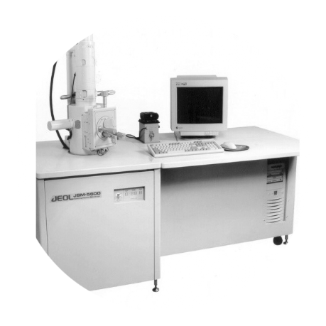

Transmission Electron Microscope (TEM) processing unit
Ultra Microtome and Histopathology
Scanning Electron Microscope (SEM) processing unit
Our Services
- Transmission Electron Microscope
- Specimen preparation as per the prescribed standards
- To support the needy people for research and diagnostic purposes.
- Timely providing of specimens for TEM and SEM
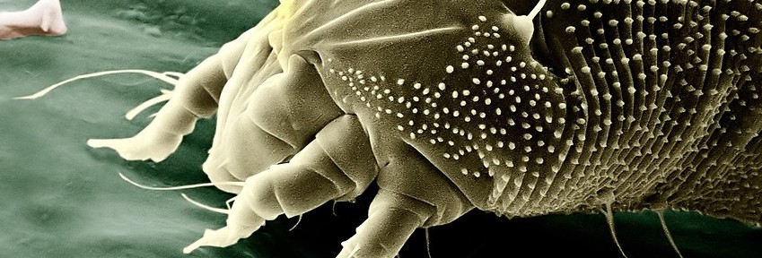
- Scanning Electron Microscope
- Transmission Electron Microscope (TEM) processing unit
- Scanning Electron Microscope (SEM) processing unit
- Ultra Microtome and Histopathology
RECENT WORKS
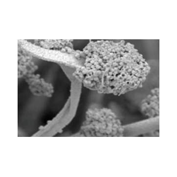
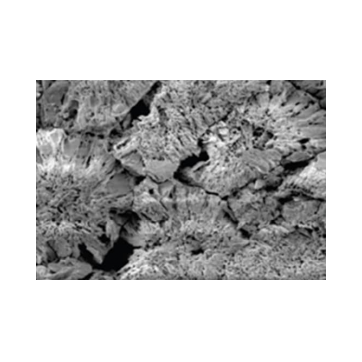
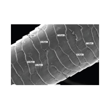
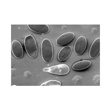
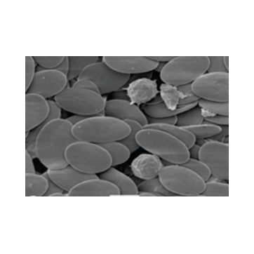
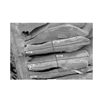
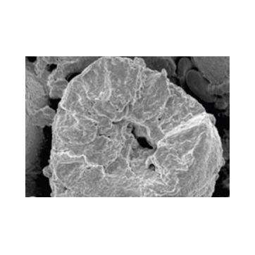
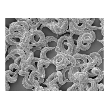
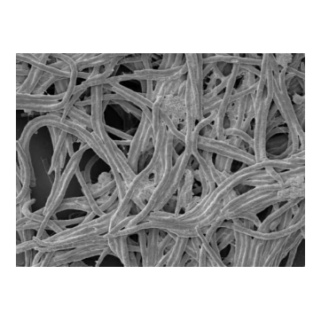
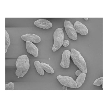
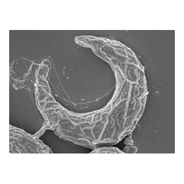
On TEM & SEM
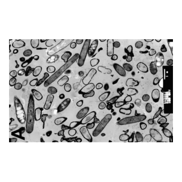
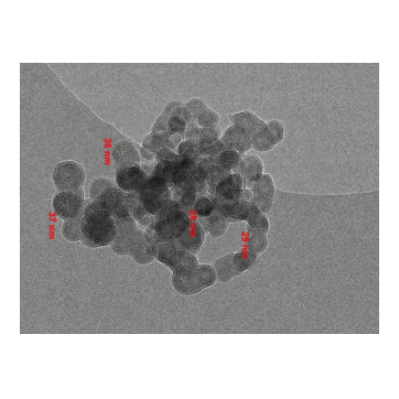
Transmission electron Micrographs (TEM) of various experimental studies

Our Clients

